| Storage | This product can be stored at -20°C for 12 months with shading light. |
| shipping | Ice bag |
| Exp date | 12 months |
| Category ID_I | Apoptosis and Cell Health Detection |
| Category ID_II | Cell Apoptosis |
| Category ID_III | TUNEL Assay Kits |
| Описание на сайте производителя | ссылка |
| Инструкция | ссылка |
| MSDS | ссылка |
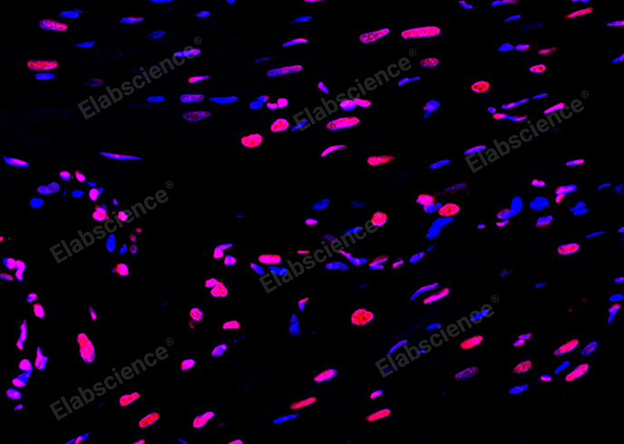
Paraffin embedded rat heart was treated with DNase I to fragment the DNA. DNA strand breaks showed intense fluorescent staining in DNase I treated sample (red). The cells were counterstained with DAPI (blue).This photo was taken by confocal microscope.
 |
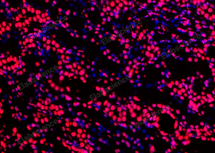
Paraffin embedded rat kidney was treated with DNase I to fragment the DNA. DNA strand breaks showed intense fluorescent staining in DNase I treated sample (red). The cells were counterstained with DAPI (blue).
 |
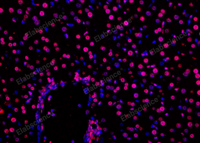
Paraffin embedded mouse liver was treated with DNase I to fragment the DNA. DNA strand breaks showed intense fluorescent staining in DNase I treated sample (red). The cells were counterstained with DAPI (blue).
 |
Fluorescence microscope analysis of camptothecin-induced apoptosis of Hela cells.
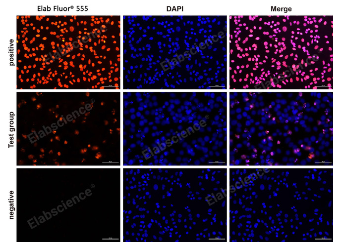 |
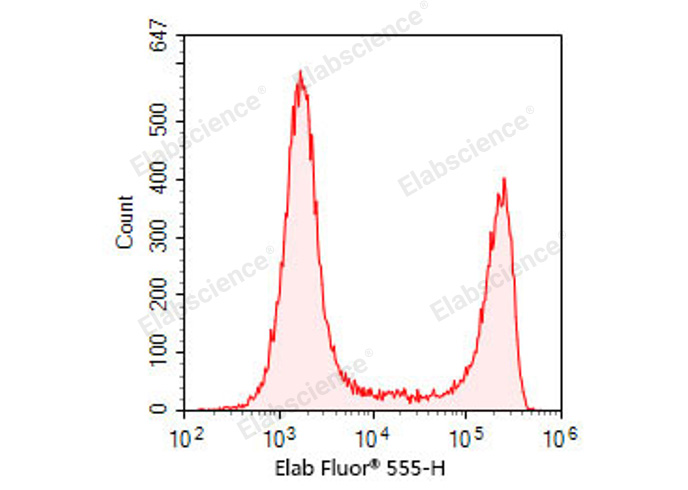
Mixed samples of normal mouse spleen cells and DNase I treated mouse spleen cells were stained.
 |
 |
| Citation link 1 | ссылка |
| Citation link 2 | ссылка |
| Citation link 3 | ссылка |
| Citation link 4 | ссылка |
| Citation link 5 | ссылка |
| Citation link 6 | ссылка |
| Citation link 7 | ссылка |
| Citation link 8 | ссылка |
| Citation link 9 | ссылка |
| Citation link 10 | ссылка |
| Citation link 11 | ссылка |
| Citation link 12 | ссылка |
| Kit Type | Apoptosis/DNA Fragmentation |
| Detection Instrument | Fluorescence Microscope |
| Tested Sample Types | Paraffin section;frozen section;cell slide |
| Assay Time | 3 hours |
| Detection Type | Fluorometric method |
| Test Principle | When cells undergo apoptosis, specific DNA endonucleases will be activated, cutting the genomic DNA between the nucleosomes. The DNA of apoptotic cells is cleaved into multimers of 180~200bp fragments, corresponding to the oligonucleosomal size. Therefore, the DNA of apoptotic cells typically migrates as a ladder of 180~200bp on an agarose gel. The exposed 3'-OH of the broken DNA can be catalyzed by Terminal Deoxynucleotidyl Transferase (TdT) with fluorescein labeled dUTP, which can be detected with fluorescence microscope. |
| | |
